


Advanced imaging of cultural heritage artefacts
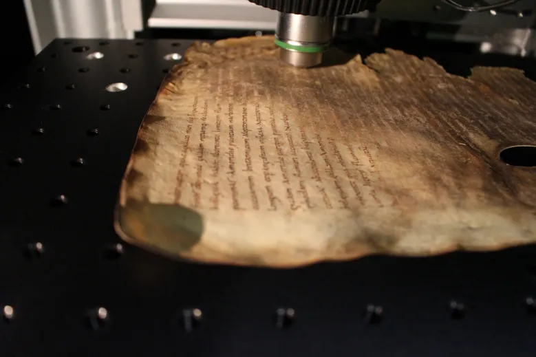 Multiphoton imaging of historical parchment
Staff
Gaël Latour / Associate Professor, Paris-Saclay University
Marie-Claire Schanne-Klein / DR CNRS
Laura Paggi / Post-doc
Internship project / Proposition de stage : Multiscale physico-chemical characterization of collagen degradation – Application to parchment
Topic
Multiphoton microscopy for the investigation of collagen-based materials
Multiphoton microscopy has revolutionized three-dimensional (3D) imaging of biological tissues by offering new modes of contrast even in unstained tissues. Since few years, some studies have shown the potential of this technique for non invasive analysis of cultural heritage artefacts. Two-photon excited fluorescence (2PEF) signals are emitted by a wide range of materials (fluorophores) in historical artifacts with specific absorption and emission fluorescence spectra. Second harmonic generation (SHG) signals are specific to dense and well aligned structures such as fibrillar collagen and cellulose.
SHG microscopy provides structural information on the 3D organization of the fibrillar collagen within parchments and other skin-based artefacts. In addition, SHG microscopy can be combined with polarimetry (analysis of the light polarization state) to measure the degree of alignment of fibrillar collagen on a sub-micrometer scale, which is closely related to the state of degradation of the parchments.
We recently applied this new methodology to the Mappa Mundi d’Albi, which is classified as “World memory” by Unesco and to historic 13th century parchment manuscripts belonging to the renowned collection at the Chartres Library.
We are developing various metrics to quantifiy the organisation and degradation of collagen and are considering using multiphoton microscopy as a tool to determine the conservation state of parchment. We are also seeking for chemical information about the degradation process by correlating the nonlinear signals with infrared spectroscopy.

Figure : (a) Imaging of a parchment before and after degradation: SHG signals (green) are emitted by fibrillar collagen, and fluorescence signals (red) appear during degradation. (b) p-SHG imaging of parchment; two types of information are extracted: the local orientation of fibrillar collagen and disorder within the focal volume.
Optical Coherence Tomography (OCT) for determining the thickness of varnish layers
Optical Coherence Tomography is another imaging technique with provides 3D images with a micrometer scale isotropic resolution. The collected signal is based on the spatial variation of the refractive index (interfaces between layers, scattering particles). This technique allows the characterization of semi-transparent layers. It is well adapted to the image of varnish layers or glazes. We have also developed image processing to automatically determine the thickness of layers. Moreover, the device is transportable and in-situ analyses are possible in conservation workshops. Several restoration projects have been carried out in recent years using OCT, including the assessment of varnish thickness on paintings and musical instruments, and the monitoring of restoration processes.
Contact : [javascript protected email address]
OCT : Access Policy, Cost Policy, and Data Policy

Figure : Left : picture of the OCT probe analyzing a painting before the conservation (Notre-Dame del Pilar, 17th century, Musée National de la Renaissance. Right : OCT images of varnish layers on the pictorial layer. The transverse views are similar to stratigraphic views.
Main publications
G. Galante, M. Vilbert, L. Desvois, D. L. Corre, L. Archambault, L. Robinet, N. Saumagne, M.-C. Schanne-Klein, and G. Latour, "Non-invasive quantitative investigation of varnish stratigraphy in historical artifacts using line-field confocal OCT," Heritage Science 14, 193 (2026) - 10.1038/s40494-026-02460-4
G. Galante, L. Robinet, S. Heu-Thao, C. Caporal, G. Latour, and M.-C. Schanne-Klein, "Intrinsic variation of the polarization-resolved SHG from collagen: Multiscale analysis and application to parchments," APL Photonics 10, 056106 (2025) - 10.1063/5.0250484
L. Robinet, S. Heu-Thao, G. Galante, G. Latour, A. Tournié, C. Daher, A. Dan, M.-C. Schanne-Klein, A. Michelin, and J. Deschaux, "The Mappa mundi of Albi: Insight into the manufacturing, life and conservation state of an 8th century world map," Journal of Cultural Heritage 74, 341-352 (2025) - 10.1016/j.culher.2025.05.013
Noninvasive quantitative assessment of collagen degradation in parchments by polarization-resolved SHG microscopy, M Schmeltz, L Robinet, S Heu-Thao, JM Sintes, C Teulon, G Ducourthial, P Mahou, MC Schanne-Klein, G Latour, Science Advances 7, eabg1090 (2021) - 10.1126/sciadv.abg1090
In-place molecular preservation of cellulose in 5,000-yeat-old archaeological textiles, C. Reynaud, M. Thoury, A. Dazzi, G. Latour, M. Scheel, J. Li, A Thomas, C. Moulhérat, A. Didier, L. Bertrand, Proc. Nat. Acad. Sci. 117, 19670-19676 (2020) - 10.1073/pnas.2004139117
Correlative nonlinear optical microscopy and infrared nanoscopy reveals collagen degradation in altered parchments, G. Latour, L. Robinet, A. Dazzi, F. Portier, A. Deniset-Besseau, and M.-C. Schanne-Klein, Sci. Rep. 6, 26344 (2016) - 10.1038/srep26344
In situ 3D characterization of historical coatings and wood using multimodal nonlinear optical microscopy, G. Latour, J.-P. Echard, M. Didier, M.-C. Schanne-Klein, Opt. Express 20, 24623 (2012) - https://doi.org/10.1364/OE.20.024623
Highlights / review articles
[French] Video Microscopie optique tridimensionnelle des matériaux anciens – La tomographie par cohérence optique (OCT) (2022)
[English] Press release A new non-invasive technique for parchment diagnosis (2021)
[French] Review La microscopie optique tridimensionnelle – De l'imagerie biomédicale à la caractérisation des objets du patrimoine (2019)
[French] Participation in the documentary "La Mappa mundi d'Albi, le monde d'hier" de Patrick Desenne (2017)
[French] Cartographier la dégradation des parchemins à l’échelle microscopique sans contact ni prélèvement, Actualités scientifiques de l’Université Paris-Sud et du CNRS (2016)
Main ongoing collaborations
- L. Robinet, S. Heu-Thao, S. Cersoy, CRC (CNRS, Museum National d’Histoire Naturelle, Ministère de la Culture, Paris)
- A. Deniset-Besseau, A. Dazzi, J. Mathurin, ICP (CNRS, Université Paris-Saclay, Orsay)
- L. Desvois (restauratrice du patrimoine, doctorante à Cergy Université)
- Institut National du Patrimoine (Aubervilliers)
- La Cambre (Bruxelles, Belgique)
- M. Thoury, IPANEMA (CNRS, Ministère de la Culture, Museum National d’Histoire Naturelle, UVSQ, Synchrotron Soleil, Saint-Aubin)
- J. Ogien, Damae Medical